Suspect Moschcowitz syndrome: how, when, why

Mantuano StefaniaMDFava AntonioMD, Costantino PaoloMD, Caterina Ceruso2 MD

1) Emergency Medicine and Trauma Surgery, Grande Ospedale Metropolitano “Bianchi Melacrino Morelli”, Reggio Calabria
2) Grande Ospedale Metropolitano “Bianchi Melacrino Morelli”, Reggio Calabria
Abstract

An 87-year-old female, who had been suffering of dyspnea and high fever for 4 days, came to our ER in yellow code. She said she had been suffering of dyspnea and orthopnoea for fifteen days even with relatively little efforts.
Laboratory data showed the presence of a mass in the right atrial cavity.
The woman was submitted to cardiologic surgery unit to be undergone to a partial neoformation removal operation.
After a histological examination, it was possible to establish that the mass was formed by a benign tumor (Lipoma).
After about thirteen occupant days in our Cardiac surgery unit, the patient moved to a centre of Cardio rehabilitation for the necessary therapies.

Keywords

dyspnea – neoformation – lipoma – cardiac surgery – Cardiopulmonary rehabilitation

Clinical Case

An 87-year-old female came into the ER (yellow code) presenting dyspnea and high fever for four days. When she was at home, she had already started a course of antibiotics for two days (Ciprofloxacin 500 mg/die). 
By her medical history, it was possible to establish she had suffered from high blood pressure
(treated with combisartan/idroclorotiazideand acetylsalicylic acid), a previous breast cancer of about thirty years ago, chronic renal failure, a previous hysterectomy. Besides, she affirmed she did not suffer from any other pathologies or allergies to medicines.
At least, she reported she had been suffering of dyspnea and orthopnoea even with relatively little efforts for the last fifteen days.
By the physical thoracic examination, the woman showed a harsh diffused breath, right breath sounds diminished. EKG showed normal sinus, with a frequency of 74 b/m and some anomalies in the ventricular repolarisation.
 
The chest X-ray was made in two orthogonal projections. The results showed a nodular opacity in the right superior pulmonary area with a diameter of about 12 mm. The cardiac shadow seemed to be lightly increased. 
Blood tests were almost all normal, except for a light chronic renal failure, already known (creatinine level 1,13mg/dl; azotaemia 94mg/dl).
A cardiological assessment was requested and the woman had to undergo an echocardiography which revealed that the left ventricle was normal with a septal hypertrophy. The pump global function was hyperkinetic. Right ventricle appeared normal for dimension and functions but there was a biatrial dilatation. Aortic diameter was normal but tricuspid aortic valve was calcified. Doppler showed a light mitral and tricuspid insufficiency. The aortic flow was modestly accelerated (V max 2,1 m/se, DP 18mmHg) and the diastolic pattern resulted altered. Exams revealed that there was a rough hiperecogen mass in the right atrial cavity, hugely planted on the back interatrial septum without engaging neither the tricuspid valve nor the inferior vena cava. The inferior vena cava could collapse breathing and, in its investigable trait, it seemed free from any pathologic intraluminal echoes.
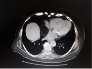
At this point, the patient was admitted to the intensive cardiologic unit care where she underwent a CT-angiography and a chest CT with contrast. Evaluating the cardiac chambers, it was possible to document the presence of a not particularly dense mass (5x5cm), with negative density values, such as for fat tissue, developing into the right atrium and comporting a reduction in volume of the same chamber. (Figure 1)

Figure 1

The ascending aorta had diameters within normal limits and it was possible to appreciate some atheromasic parietal calcifications at the aortic arch and the descending thoracic aorta. There was neither pericardial effusion, nor airway obstruction.
The patient underwent a coronary catheterization that pointed out that the coronary anatomy with left dominance did not present any significant lesions and there was an abnormal origin of the right coronary from the circumflex branch.
After five days into the intensive unit care, the patient moved into the Cardiology Unit where she had a partial removal of the neoformation on extracorporeal circulation. The patient underwent a coronary catheterization that pointed out that the coronary anatomy with left dominance did not present any significant lesions and there was an abnormal origin of the right coronary from the circumflex branch. During the operation, the patient had a median sternotomy and a pericardiotomy. I.V. Heparinwas given the patient, and it was made an ascendant and bicameral incannulation. The operation was made on extracorporeal circulation. The surgeons were able to clamp the aorta, instilling cardioplegia into the aortic bulb while the heart was asystolic. The surgeons clamped the pulmonary trunk, and they practised an incision in the right atrium during the extracorporeal circulation. It was possible to see a huge mass of about 6 cm extended to the interatrial septum and to the free walled atrium. The mass seemed to be a lipoma and it was partially removed.
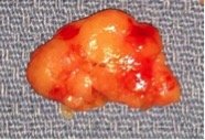
The deep and comprehensive extension of the mass, without a cleavage, made impossible its total removal but, anyway, the surgeons were able to free, as far as possible, the two cava veins and the coronary sinus. The tricuspid valve was not obstructed. A sample was sent to be analysed. The extemporaneous biopsy before, and a second biopsy later, confirmed the diagnosis of lipoma. (Figure 2)

Figure 2

The right atriotomy was sutured with prolene 4/0 and a heterologous pericardial patch and glue were applied on the suture (Periguard 8×14 cm). The air was purified and the surgeons declamped the pulmonary artery and the aorta. The pulse was back to normal after a 20Jx1 shock and the general circulation was reestablished without any kind of problems through the use of inotropes LED. Decannulation was made, the haemostasis was made through the use of protamine. Two drainages were applied: the first was pericardial, the second mediastinal. The right ventricle was sutured using polypropylene wire. The pericardium was closed over the aorta. The sternum was symmetrically pulled over with metallic wires and the skin was sutured.
The clinical course was regular, without any relevant pathological clinic event. The following extubation of the patient was made in total security by ensuring regular breathing acts, even thanks to an intense breathing physiotherapy. 
The patient moved into the Recovery Unit the day after the surgical procedure. She started to move regularly with the help of the physiotherapists.
The following instrumental tests (chest X-ray and an EKG) showed the presence of a residue of the mass into the right atrium headed toward the atrial septum. The EKG revealed a tricuspid valve insufficiency of medium degree and a blood systolic pressure within the pulmonary artery of about 60mmHG.
After about thirteen days in the cardiac division of our centre, the patient moved into a cardiac rehabilitation unit for the necessary following therapies.

Short historical hints

The primary tumors of the heart in adulthood can be benign or malignant. They have a variable incidence, the frequency of primary cardiac tumors is approximately 0.02%, corresponding to 200 tumors in 1 million autopsies1. The benign tumors are more common than the malignant, at a 3:1 ratio.

The most common benign tumors, ordered by frequency, are the myxoma, the lipoma, the Papillary Fibroelastoma, the rhabdomyoma, the fibroma, the haemangioma, the teratoma, the mesothelioma of the atrioventricular node, the tumor of the granular cells, the neurofibroma and the Lymphangioma2.
The most common malignant tumors are: the angiosarcoma, the rhabdomyosarcoma, the mesothelioma, the fibrosarcoma, the malignant lymphoma, the osteosarcoma, the thymoma, the neurogenic sarcoma, the leiomyosarcoma, the liposarcoma, the synovial sarcoma3.
The tumors of the heart are very similar to those of the striated muscles. Symptom patterns depend from how much the heart is involved. For this reason, it is possible to have pleural and pericardial pain, dyspnea or angina. We can also occasionally observe systemic symptoms, such as embolus, cachexia and syncope.
The most common clinical signs are the flexure of the fingertips, fever, flushing, pericarditis, arrhythmias, anomalies in leading. In some case, the situation could be worse, with cases of heart attack, heart failure, reduction of atrial cavities, dysfunction or obstruction of the valves.
The most frequently used diagnostic techniques are the echocardiography, the computer tomography, the magnetic resonance and the imaging through radionuclides. 
The treatment is exclusively surgical, considering also the heart transplantation. 
The malignant tumors need a supportive therapy such as chemotherapy and radiation4.

Bibliography

  1. Reynen K. Frequency of primary tumors of the heartAm J Cardiol 1996; 77: 107. 
  2. Grebenc ML, Rosado de Christenson ML, Burke AP, Green CE, Galvin JR. Primary cardiac and pericardial neoplasms: radiologic-pathologic correlation. Radiographics 2000; 20: 1073-103.
  3. S Tremosini et al – Sarcoma primitivo del cuore. Descrizione di un caso. G Ital Cardiol 2007; 8 (3): 196-200
  4. Sarjeant JM, Butany J, Cusimano RJ. Cancer of the heart: epidemiology and management of primary tumors and metastases. Am J Cardiovasc Drugs 2003; 3: 407-21.