- Marta Manghetti
- Brief Report and Case Report
Isolated spontaneous dissection of the celiac artery: a case report
- 2/2018-Luglio
- ISSN 2532-1285
- https://doi.org/10.23832/ITJEM.2018.022
Marta Manghetti*, Claudia Gianni**, Pietro Bemi**, Laura Bassani**, Fabiana Frosini*
*S.C. Pronto Soccorso e Medicina d’Urgenza Ospedale San Luca, Lucca. Usl Toscana Nord Ovest
**U.O.C. Diagnostica per Immagini Lucca e VDS. Usl Toscana Nord Ovest
Abstract
Arterial dissection is defined as the cleavage of the arterial wall by an intramural hematoma. Reports of spontaneous isolated visceral artery dissection are rare.
A 46-year-old male presents to the emergency department with epigastric pain, a common symptom. A Computed Tomography (CT) Angiography of the thoracoabdominal aorta was applied and dissection of celiac, splenic and hepatic artery was determined. A conservative therapeutic approach was preferred and the patient was discharged with medical therapy.
Spontaneous isolated celiac arterial dissections have to be considered in the differential diagnosis of the epigastric pain. Contrast-enhanced CT angiography examination is the main method in the diagnosis.
Keywords
Celiac artery; dissection; epigastric pain; angiography; emergency depertment.
Introduction
Case Report
A 46-year-old previously healthy male presented to our Emergency Department for acute abdominal pain. He presented pain during the previous 3-days. The pain was localized in epigastrium and left–upper abdominal quadrant and was non-radiating. The day before the arrival at the Emergency Department, he vomited. He denied diarrhea, fevers, bright red blood per rectum, melena or rigors. The patient had no history of hypertension and his vital parameters were normal: blood pressure 150/80 mmHg, heart rate 78 bpm, 15 breath/min, body temperature 36°C. His medical history included tonsillectomy and varicocele surgery.
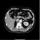
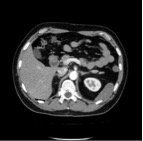
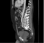
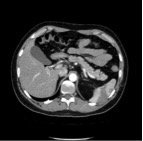
At presentation, physical examination revealed mild epigastric tenderness without guarding or rebound, with normoactive bowel sounds. Biochemical test demonstrated only aspecific leucocitosis. The elettrocardiogram showed normal sinus rhythm. Abdominal radiography and sonographyc examination revealed normal pattern. We administered intravenous symptomatic therapy without success. The patient underwent an abdominal CT angiography examination with a 64-MDCT scanner (Toshiba Aquilion CX) receiving 110 mL of iomeprol (400 mg I/mL) injected at a flow rate of 3,5 mL/s. Original axial thin-slice images (Fig. 1 and Fig.2) and reconstructed sagittal (Fig.3) revealed a dissection of the celiac artery with a flap that originated from the aortic ostium, extended past the origin of the left gastric artery and into the bifurcation of the artery into the splenic and common hepatic artery. There was no aneurysmatic dilatation of the celiac artery. The true lumen was patent with contrast seen in the hepatic, left gastric and splenic arteries. There were splenic infarction findings; no other visceral damage was detected (Fig.4).
Figure 1. Origin of the celiac artery dissection starting from the aortic ostium
Figure 2. Proximal celiac artery patent only in true lumen
Figure 3. Sagittal reconstruction showing the emergence from the aorta of the celiac artery with medium contrast only within the true lumen
Figure 4. Dissection of the hepatic artery and splenic artery. Splenic infarctions
After 7 days the patient was discharged with a medical therapy and planned follow-up CT examinations.
Discussion
The initial symptom in most patients is acute or chronic epigastric or hypochondrial pain. Patients with ruptured aneurysms may present acutely with bleeding. Chronic dissection has symptoms such as postprandial abdominal pain, intestinal angina and weight loss. Most patients with celiac artery dissection are asymptomatic possibly due to the lack of small bowel involvement (4). The average age of patients is approximately 55 years (5). Visceral artery dissections are 5 times more common in males than in females. (5) Risk factors are described as hypertension, cystic medial necrosis, abdominal aortic aneurysm, fibro-muscular dysplasia, trauma, pregnancy, connective tissue disorders (6). No definite causes have been found in many cases. The natural history of this condition has not yet been fully clarified (7). The histology has shown that the dissection occurs between the intima and the external elastic layer, whereas in dissection of the aorta the cleavage plane is between the first and the second part of the intima (1).
The diagnosis of artery dissection largely depends on radiological findings (6).
CT angiography is the preferred method for the evaluation of vascular structures and related pathologies. It is as accurate as conventional angiography with less morbidity, and it is less expensive. The CT scan is considered the primary technique for diagnosing celiac artery dissection. The CT scan can provide high quality images of the dissection site and provide information about the extent of the lesion, presence of aneurysm formation or intramural hematomas (8).
There is no consensus about the more appropriate treatment. Open surgery, endovascular surgery, and interventional radiology are invasive options for the management of visceral artery dissections. Conservative treatments including anticoagulants or anti-platelet agents for 3-6 months with a target INR of 2-3, antihypertensive therapy, anti-inflammatory drugs or steroids. The subendothelial injury can trigger thrombosis. Strict blood pressure control might prevent propagation of the dissection (9).
Conclusions
Spontaneous visceral artery dissection has to be taken in consideration in the differential diagnosis of epigastric pain in the emergency department. The unavailability or the rarity of angiographic CT examinations could underestimate this rare disease. Although extremely rare, patients presenting with epigastric pain and risk of vasculopathy should be considered for isolated spontaneous visceral artery dissection. The improvements in the use of contrast-enhanced CT in emergency department could make a easier diagnosis. Early recognition and definitive therapy lead to a favorable outcome. Surgical intervention may be considered for significant bleeding or signs of intestinal ischemia. The reported cases are too few and the follow-up periods too brief to yield definitive conclusions (10).
References
(2) Aydin S. et al: Spontaneous Isolated Celiac Artery and Superior Mesenteric Artery Dissections: a rare case. Pol J Radiol, 2015: 80: 470-472.
(4) Vaidya S. and Dighe M.: Spontaneous celiac artery dissection and its management .J Radiol Case Rep. 2010; 4(4): 30-33.
(6) Ji Yeon Lim et al: Unusual presentation and treatment of isolated spontaneous gastric artery dissection. Clin Exp Emerg Med 2016; 3(2): 112-115.
(8) Subhas G. et al: Spontaneous isolate superior mesenteric artery dissection: a case report and literature review with management algorithm. Ann Vasc Surg 2009; 23 (6): 788-98.
(10) Melnychuk E. and Strony R.: Spontaneous Isolated Visceral Artery Dissection in a Middle Aged Male. Case Reports in Emergency Medicine; 2017, article ID 3704348, 3 pages.
Referees
Prof. Mauro Ferrari – Direttore Dipartimento Cardio Toraco Vascolare – U.O. Chirurgia Vascolare Ospedale Cisanello, Azienda Ospedaliera Universitaria Pisana. E.mail: m.ferrari@med.unipi.it
Prof. Davide Caramella – Direttore U.O. Radiodiagnostica 3 Ospedale Cisanello, Azienda Ospedaliera Universitaria Pisana. E.mail: davide.caramella@med.unipi.it
Dr Alberto Conti – Responsabile Area Emergenza Urgenza Usl nordovest Toscana, Direttore Pronto Soccorso Massa Carrara, USL nordovest Toscana. E.mail: alberto.conti@uslnordovest.toscana.it


