Detection of left atrial compression by focused cardiac ultrasonography in a patient with worsening dyspnea

Alfonso Sforza1,2, Costantino Mancusi1, Maria Viviana Carlino1,2, Giovanni Porta2, Venere delli Paoli2, Giuliano De Stefano1, Giovanni de Simone1, Fiorella Paladino2
1. Hypertension Research Center, UOC Emergency Medicine, Federico II University Hospital, Naples.
2. Emergency Department, Cardarelli Hospital, Naples.

Case Presentation

 A 62-year-old man with history of gastric cancer presented to our Emergency Department (ED) with progressive dyspnea and reduced exercise tolerance during last weeks. At admission blood pressure was 110/70 mmHg, heart rate 70 beats/minute regular, oxygen saturation was 98% (FiO2 21%) with mild tachypnea (22 Breaths per minutes) and he was afebrile. Chest examination revealed decreased vesicular murmur in the base of left lung. Cardiovascular examination revealed a normal cardiac rhythm, no murmurs, normal peripheral pulses and no edema. Arterial blood gas analysis on room air revealed respiratory alkalosis with mild hypoxemia. The ECG showed sinus rhythm with normal AV conduction, normal axis and QT interval.
Results of blood tests showed a normal white blood cell count (6.660 cells per mm3), with renal and liver function test and serum electrolytes within the reference limits.
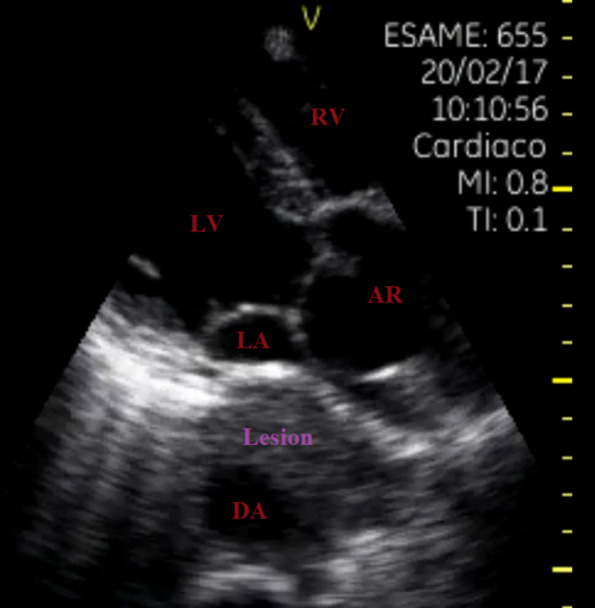
Focused cardiac ultrasonography (FoCUS) showed normal left ventricular (LV) size and function with annuloaortic ectasia and compression of left atrium (LA) due to a large mass englobing the descending aorta (DA) (Figure 1). Lung ultrasonography (LUS) revealed bilateral A-line pattern.

Figure 1. Focused cardiac ultrasonography: annuloaortic ectasia and compression of left atrium due to a large lesion englobing the descending aorta. Legend RV: right ventricle, LV: left ventricle, LA: left atrium, AR: aortic root, DA: descending aorta, Lesion: mass of unknown origin.
 
To discriminate between a descending aortic aneurism with endo-luminal thrombus and a mass inglobating DA, the patient underwent chest CT scan.
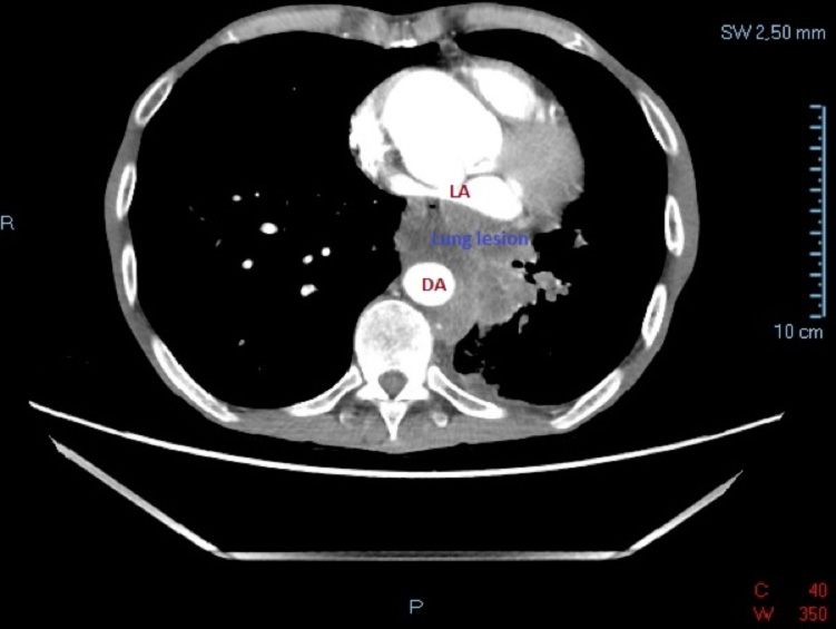
Contrast-enhanced CT scan showed ectasia of aortic root and ascending aorta and a large lesion (8x9x11cm) in the left lung suggestive of Lung Cancer (LC) inglobating DA and compressing LA (Figure 2).


Figure 2. Contrast-enhanced CT scan: large lesion suggestive of lung cancer inglobating descending aorta and compressing left atrium. Legend LA: left atrium, DA: descending aorta.
 

Discussion

LA compression by extra-cardiac structures is a rare cause of dyspnea or reduced exercise tolerance and can easily be detected by transthoracic echocardiography (TTE) (1) . Compression of LA reduces the chamber volume causing low cardiac output or even obstructive shock. Furthermore, pulmonary venous pressure increase can be cause of dyspnea, reduced exercise tolerance or even pulmonary edema (1).
Extrinsic LA compression can be caused by: gastrointestinal structures, which are the most common, mediastinal structures, aorta, intra-pericardial structures and pulmonary structures (especially tumor masses or bronchogenic cysts) (2).
Aneurysm of the aortic root and descending aorta, easily recognizable  by TTE, can cause LA compression, configuring a life-threatening condition (3).
We describe a case of LC extended to DA and compressing LA. Considering the presence of annuloaortic ectasia, to discriminate between descending aortic aneurysm in the presence of endo-luminal thrombus and a mass inglobating DA, the patient underwent immediatechest CT scan with contrast (4).
FoCUS is a reliable tool in the management of patients with acute dyspnea giving the opportunity to assess cardiac structure and function immediately after the first clinical work-up in ED (5). Although the definite diagnosis is usually confirmed by other imaging modalities, it is convincible that FoCUS is the first tool to detect LA compression in patients presenting to the ED for dyspnea.
 

Financial/nonfinancial disclosures

None declared.

References

  1. Walpot J, A. B. (J Am Soc Echocardiogr. 2007 Oct;20(10):1220.e4-6. Epub 2007 Jun 20). Left atrial compression by dissecting aneurysm of the ascending aorta.
  2. van Rooijen JM, v. d. (Eur J Echocardiogr. 2008 Sep;9(5):661-4.). Left atrial impression: a sign of extra-cardiac pathology.
  3. Bouzas-Mosquera A, B.-C. E.-V.-B. (Rev Esp Cardiol. 2008 Jan;61(1):98-9). Left atrial obliteration due to an aortic aneurysm secondary to chronic aortic dissection.
  4. Mahajan K, A. S. (BMJ Case Rep. 2016 Jan 28;2016. pii: bcr2015214213). Left atrial impression by a dilated oesophagus in a patient with limited cutaneous systemic sclerosis.
  5. Sforza A, M. C. (Cardiovasc Ultrasound. 2017 Jun 19;15(1):16). Diagnostic performance of multi-organ ultrasound with pocket-sized device in the management of acute dyspnea.