- Antonio Pagano
- Brief Report and Case Report
A case of cardiac perforation
- 3/2017-Ottobre
- ISSN 2532-1285
- https://doi.org/10.23832/ITJEM.2017.022

Abstract
Cardiac perforation by permanent pacemaker lead is a rare but possible event. Usually it occurs early, but less frequently it is a late onset complication. We report a case of delayed perforation by passive fixation lead, two weeks after the implantation, in a patient with no comorbidities. The strumental and clinical integrated approach allows rapid framing even complex clinical cases.
Introduction
Cardiac perforation by permanent pacemaker lead is a rare but possible event. Usually it occurs early, but less frequently it is a late onset complication. We report a case of delayed perforation. The diagnosis was quickly with the integrated approach. Point of care ultrasound is fundamental in the setting of emergency medicine.
Case description
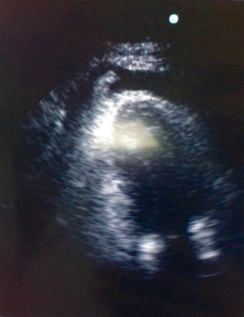
Fig 1: ultrasound image of pacemaker lead dislocation
Discussion
Cardiac perforation after pacemaker implantation is rare complications (0,1-0,8%) (1). Perforations occurring within 24 h after implantation are labeled as acute; those occurring within one month after implantation are subacute while perforations which occur after one month are labeled as chronic. Risk factors for lead perforation include patient characteristics such as female sex, age, small body habitus, thin heart walls; concomitant therapies such as steroids or anticoagulants; implant techniques; and the design characteristics of the lead (2). The symptoms depend by location of pacemaker lead: dyspnea, chest pain, emodinamyc instability if developed a cardiac
tamponade.
Management depends on patients’ hemodynamic status. In most patients, percutaneous lead extraction is a safe and effective management approach (3). Emergent surgical management is required if the patient is hemodynamically unstable or if the patient has a large pericardial effusion where tamponade may be imminent or a large pleural effusion with respiratory impairment is present or imminent. No consensus exists regarding the appropriate management of lead perforation in stable patients without symptoms or the management of chronic lead perforation without pacemaker malfunction.
References
- Lead Perforation: Incidence in Registries. Pacing and Clinical Electrophysiology. Volume 31, Issue 1, pages 13–15, January 2008
- Case of early right ventricular pacing lead perforation and review of the literature. World J Clin Cases. Jun 16, 2014; 2(6): 206-208
- Incidence, management, and prevention of right ventricular perforation by pacemaker and implantable cardioverter defibrillator leads. Pacing Clin Electrophysiol. 2014 Dec;37(12):1602-9


