Late-onset deficit of ornithine transcarbamylase: a case report and review of the literature

Pietro Pozzessere1, Michela Nardacci2, Mariangela Portaluri2, Serafina Schiraldi2, Vito Procacci1

1) Department of Emergency Medicine and Surgery “Azienda Ospedaliera Consorziale Policlinico” of Bari, Italy
2) Postgraduate school in Emergency Medicine University of Bari Medical School Bari, Italy
Abstract

Introduction

Hyperammonaemia can occur if there is an increased production of ammonia or when ammonia detoxification is affected. Ammonia crosses the blood-brain barrier and it is toxic because it may result in irreversible brain damage if not early and thoroughly treated. Causes of non-hepatic hyperammonaemic coma are not common while the urea cycle disorders are frequent. Ornithine transcarbamylase (OTC) deficiency is the most common of the urea cycle disorders, which can occur both in children, and less commonly, in adults. It is acquired as X-linked trait or a new mutation. In case of late-onset its recognition is difficult.

Case report

We report the case of a previously healthy 21 year-old white man admitted to our Emergency Room for a new-onset of slurred speech and nausea during the last few hours. He had inhaled spray paint  two days before. During the examination, he later showed aggressiveness and finally a rapid decline of his mental status leading to coma after a few hours. A specific intravenous therapy, based on arginine and sodium benzoate was administered to our patient.
Due to his persistently elevated serum ammonia levels, also hemodialysis (CVVHDF) was initiated. After more prolonged sessions, his level of consciousness did not get better also during transient progressive reduction of ammonia levels. Four days after, the patient was declared dead. A sequence analysis of OTC gene showed hemizygosis for the pathological variant known as c. 392T>Cp. (Leu131Ser) of OCT gene.

Conclusion

It is necessary to always consider ammonia levels in adult patients affected by unexplained coma. The correlation between plasma ammonium concentration and hepatic encephalopathy is not consistent, thus suggesting that other factors are important in producing brain edema. Case reports like this are important because they show that the late phenotypic expression of OCT deficiency carriers is the result of the interactions between environmental conditions and genetic anomalies.

Keywords

Hyperammonaemia, Ornithine transcarbamylase deficiency, late-onset defect of urea cycle

Introduction

Plasma concentrations of ammonia in the systemic circulation are normally very low (<40 µmol/L in adults, <100µmol/L in newborns)1.
Most of the systemic ammonia pool is originally generated in the gastrointestinal tract with the cleavage of urea in ammonia and carbonate by urease producing bacteria in the colon2
The mucosa of the small intestine produces a large amount of phosphate-activated glutaminase (PAG) and little glutamine synthetase (GS). Both enzymes incorporate ammonia into glutamine which is released into the circulation. Glutamine is metabolized by PAG to ammonia and glutamate, which is oxidized to citrulline. Ammonia is carried to the liver and is converted to urea in the periportal hepatocytes, while citrulline passes into the blood and is incorporated into arginine in the kidneys3. Hepatocellular dysfunction results impaired clearance of ammonium by the liver7.
When the urea cycle does not control the ammonia load, hyperammonaemia appears. Ammonia crosses the blood-brain barrier and it is toxic only for the brain. The astrocytes contain 80% of brain GS and can incorporate ammonia into glutamine. Hyperammonaemia may result in irreversible brain damage if not early and thoroughly treated6, infact patients died from acute hyperammonaemia have gross cerebral edema and the astrocytes are swollen4-5.
Without effective treatment, hyperammonaemia causes clinical symptoms such as vomiting and after, somnolence, disorientation until coma and death. Occasionally the onset is not specific and it is attributed to neurological and psychiatric disorders or seizures.
The correlation between plasma ammonium concentration and hepatic encephalopathy is not consistent, thus suggesting that other factors are important in producing brain edema8. The main prognostic factors are peak ammonia concentrations and duration of coma10.
Severe symptomatic hyperammonaemia which is not due to liver diseases is not common.
Hyperammonaemia can occur if there is an increased production of ammonia  (e.g. infections, drugs ecc.) or when ammonia detoxification is affected (e.g. inherited defects of the urea cycle, portosystemic shunts ecc.)9.
 
  • Inherited dfects of the urea cycle
  • Secondary urea cycle disturbance     1 – Inherited: amino acid transporter defects     
                                                                                    2 – Acquired: nutritional arginine deficiency  
  • Inherited defects of fatty acid oxidation
  • Inherited organic acid disorders
  • Hypovolaemic shock, congestive cardiac failure
  • Circulatory abnormalities with portosystemic shunting
  • Medications; anticonvulsants: sodiumvalproate, topiramate (carbamazepine?); chemotherapeutic drugs: asparaginase, 5-fluorouracil; salicylates (with other predisposing factors)
  • Haematological malignancies: multiple myeloma, leukaemia
  • Urinary infection with urease-producing bacteria
  • Excessive amino acid load/increased catabolism: gastrointestinal haemorrhage, glycine irrigation, cachexia+high protein feeds
Table 1. Causes of non-hepatic hyperammonaemia in adults
 
Hyperammonaemia is classified as primary hyperammonaemia when it is directly affected by a defect of any of the involved enzymes or transporters of the urea cycle11.
For all enzyme, as well as for the two antiporters, there are known disorders when any of the encoding genes carries a defect12.
When the urea cycle is inhibited by accumulating metabolites or substrate deficiencies, the resulting hyperammonaemia is called secondary13
Among all these disorders the most relevant group is the one of organic acidemia sas methylmalonic acidemias or isovaleric acidemias, in which there is an inhibitory action of specific metabolites on the function of N-acetyl glutamatesynthetase (NGAS) or Carbamoylphosphate synthetase(CPS1)14.
The metabolites of valproic acid having an inhibitory reversible effect on NAGS activity15.
Moreover, many substrate deficiencies may impair the urea cycle function through the inhibition of NAGS (lysinuric protein intolerance, fatty acid oxidation defects or defects of the carnitine cycle16).
Finally, there are forms of acquired hyperammonaemia.
The most frequent are  the ones due to an increased production of ammonia (urinary tract infections with urease-production organism, e. g. Proteus Mirabilis and some Klebiella species)17, in subjects taking drugs, as L-Asparaginase16 or in a few patients receiving parental nutrition (no further reports in literature after 2001)17.
Hyperammonaemia may be due to a inherited defect of glutamine synthetase (GS)18 or acquired GS deficiency after orthotopic lung- or bone-marrow transplantations19.
It was described in portosystemic shunting between portal vein and inferior vena cava where intestinal blood  is deviated  into the systemic circulation20.
In transient hyperammonaemia of the newborn (THAN) an open ductus venosus probably leads to a shifting of blood into the inferior vena cava21.
Taking  into account all these reasons we have to think of hyperammonaemia in all patients with encephalopathy  even if they have no liver diseases. 
To this end, we hereby describe a report of fatal coma in a young adult with a congenital late-onset OCT deficiency.

Case Report

A 21-years-old white man, previously healthy, was admmittedto the Emergency Room following a new-onset in a few hours of slurred speech and nausea.

The patient denied abuse of illegal substances during the days before. He was accompanied by his parents. They assumed that the manifestations were due to inhalation of spray paint (alkyd-urethane enamel) happened two days before. 
In the morning he had  been already examined for sore throat, headache and emesis he had been experiencing for two days, without any associated symptoms. He was discharged with a diagnosis of suspected intoxication by inhalant chemicals. 
Moreover, his parents reported that he had been showing a solitary  behavior for two years, and followed a disordered diet, rich in proteins and lipids and that he had been using  drugs (cannabis) up to two years before. No family history of epilepsy and neuropsychiatric disorders was reported.
On admission he was afebrile, his vital signs were normal, except for a sinus tachycardia (heart rate was 120 bpm). The neurological examination only revealed an ideo-motor slowdown. His Glasgow Coma Scale was 15. 
Initial laboratory data showed normal leukocyte count with neutrophilia (85%), mixed hyperbilirubinemia (total bilirubin 4 mg/dL, non-conjugated 3.67 mg/dL), abnormal coagulation panel (PT INR 1.44, aPTT Ratio 1.24) and low fibrinogen levels (98 mg/dl, v.n. >200 mg/dl). The toxicological panel was normal (ethanol, amphetamines, barbiturates, benzodiazepine, cannabinoid, cocaine, methadone, opioid). Blood gas revealed both respiratory and metabolic alkalosis. The computed tomography (CT) scan of the brain without contrast enhancement was also normal. An ultrasound scan with Doppler of abdomen revealed normal hepatic parenchyma.
Over the first hour of admission, he began to cry, showing restlessness and aggressiveness; during the psychiatric examination 1 vial of intramuscular carbamazepine was administered to the patient showing a progressive lethargy. 
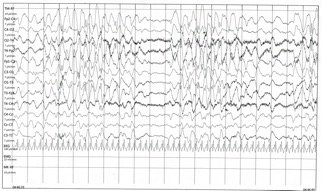
For this reason, the patient  underwent a prolonged electroencephalography (EEG) that showed a slow background activity, interrupted by short pauses without electrical activity. 
 

Figure 1. This electroencephaloghraphy portion of long-term tracing demonstrates background slowing in the range of 3HZ with brief intervals characterized by reduction of electric activity

 
Further diagnostic examinations such as magnetic resonance angiography (MRA) of the brain and total body CT were all normal. Considering his hypertonic response to nociception and the  bilateral signs of decerebrate rigidity, he was intubated for airway protection and admitted into intensive care unit. In order to prevent seizures, 1 vial of valproic acid was administered, after replaced with levetiracetam.
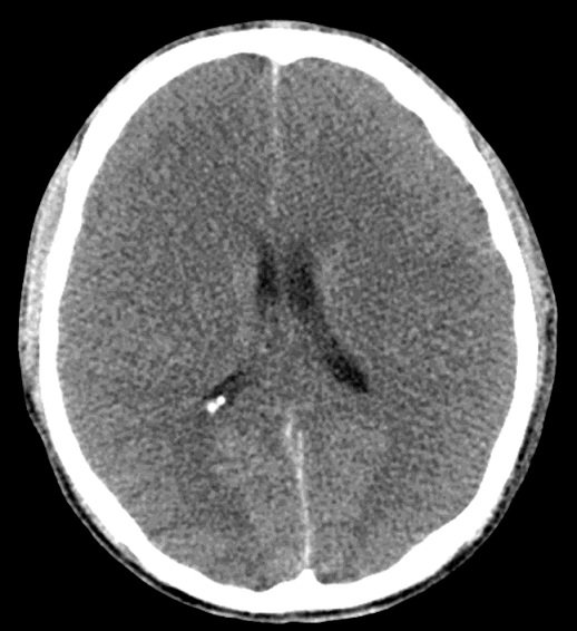
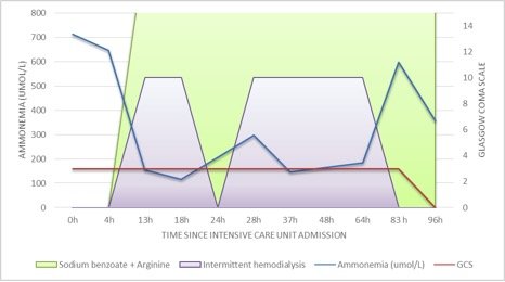
Later blood tests showed high level of ammonia (714 µmol/L). Consequently, a specific intravenous therapy, based on arginine and sodium benzoate, with an addition of protein-free nutritional support, was administered to our patient. Another brain CT was performed after 24h showing diffuse cerebral edema. 
Figure 2. Computed tomography brain scan without contrast showing a generalized loss of the supratentorial grey-white matter differentiation with effacement of sulci indicating diffuse cerebral edema with size reduction of ventricular system

Urea cycle disorder was strongly suspected and so hemodialysis (CVVHDF) was initiated as well as plasma and urine amino acid analysis and urine organic acid quantitation were performed. After the second hemodialysis session we witnessed a transient progressive reduction of ammonia level without any observable improvement in his level of consciousness.
The blood test showed high level of glutamine, ornithine, arginine, lysine and normal level of citrulline, while the urine test revealed elevated levels of orotic acid (4,4 uM/M creatinine; v.n.<0,29). A liver biopsy was performed for genetic testing. The transcranic color doppler ultrasound, performed two days after the admission in ICU, certified absence of cerebral flow and four days later the patient was declared dead. The genetic testing was performed post-mortem by the CoNVaDING tool and confirmed by multiplex ligation-dependent probeamplification (MLPA).
The sequence analysis of OTC gene showed hemizygosis for pathological variant known as c.392T>Cp.(Leu131Ser) of OCT gene.
Figure 3. Diagram showing a timeline of the measured ammonia level in the serum, the level of consciousness (as GCS) in relation to ammonia scavenger therapy (arginine+sodium benzoate) and intermittent hemodialysis

Discussion

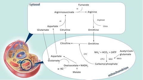
The urea cycle is the main way through which the ammonium and nitrogen in excess bound to the amino groups are converted into urea and then excreted in the urine. It is the result of 6 consecutive reactions, three mitochondrial and three in the cytosol, and at the end of each cycle two nitrogen atoms are converted into urea, one that comes from ammonium and another from the amino acid pool, with the alanine as the main component, both channelled through aspartate.

Two transport proteins (vitrine and ornithine transporter-1) and an enzyme cofactor (N-acetyl glutamatesynthetase or NGAS) are also involved in the cycle22,23.
The 5 that catalyze the enzymatic reactions are:
Carbamoylphosphate synthetase (CPS-1), Ornithinetranscarboxylase (OTC), argininosuccinatesynthetase (ASS), argininosuccinatelyase (ASL) and Arginase (ARG).
These enzymes are present in the liver, intestines and kidneys but only in the liver reach levels capable of supporting physiological ureogenesis.
In order to work, the CPS-1 has absolute necessity of N-acetylglutamate (NAG), an allosteric activator produced by the reaction between acetyl-CoA and glutamate catalyzed by the enzyme NAG synthetase (NGAS).
The short-term regulation of the cycle is achieved through the action of the NAG on the CPS-1 and reflects the availability of acetyl-CoA, glutamate and arginine, the latter activator of the NAGS25.
The long-term control, on the other hand, is obtained through an altered enzymatic production, probably through the action of transcription factors and hormonal regulation.
A defect or absence of one of the enzymes or one of the 2 substrate transporters or co-factors determines hyperammonaemia26,27.
Figure 4. The urea cycle. NAG, N-acetylglutamate; NAGS, N-acetylglutamate synthetase; CPS I, carbamyl phosphate I; OTC, ornithne transcarbamylase; AS, argininosuccinic acid synthetase; AL, argininosuccinic acid lyase;  ɑ -KG, 2-oxoglutaric acid; 1, mitochondrial ornithine transporter (ORNT 1); 2, aspartate-glutamate carrier (CITRIN)
 
The incidence of urea cycle disorders in the US is one in 35,000 new-borns, corresponding to 113 new patients a year considering all age groups. Considering the same incidence, 149 new cases are expected each year in European countries. 30 new-borns a year in the USA present hyperammonaemia (26% of cases).
There are 9 different genetic defects. The OCTD deficiency is the most frequent anomaly and its incidence is 1 in 56,500 new-borns/year (57-62% depending on the national registries (Urea Cycle Disorders Consortium (UCDC), National Urea Cycle Disorders Foundation (NUCDF) e European Registry and Network for Intoxication Type Metabolic Diseases (E-IMD)28.
All anomalies are transmitted in an autosomal recessive manner except for OCTD which is the only defect linked to sex (X-linked): the human OCT gene is located on the short arm of the X chromosome in the Xp21.1 band29,30.
The onset of the symptomatology can occur within the first month of life (neonatal onset or NO) or after the first month of life (late onset or LO) with different mortality rates: 24% in the first and about 11% in the second. The morbidity in both cases is instead high. Researchers hypothesize that complete OCT deficiency is associated with neonatal onset while hemizygous forms are associated with late onset. Heterozygous women are asymptomatic in 60-80% of cases; the remaining 20-40% behave like men with partial defect, even if they have a postponed onset on average31,32.
LO diseases in male hemizygous are caused by point mutations that result in lower enzymatic activity, a destabilized protein, or decreased affinity for the substrate. In most women patients and in some men the mutation may appear “de novo” and the mother is not a carrier33.
The asymptomatic forms naturally have significant clinical repercussions especially on family planning. Our patient was a 21-year-old male who had never shown hyperammonaemia in the past, nor similar stories among close relatives. He came to the emergency room for episodes of vomiting in the previous two days and the appearance of a worsening ideomotor slowdown.
On arrival, he presented mild respiratory alkalosis, normal transaminase and increased bilirubinaemia, lengthening of INR and hypofibrinogenaemia.  
Hyperventilation and the consequent respiratory alkalosis generally precede or accompany acute metabolic crisis, cerebral oedema and the typical coma of hyperammonaemia.
Respiratory alkalosis is the anomaly of the predominant acid-base balance in patients with various enzymatic deficits in the urea cycle24,29,34,.
Patients with symptomatic OCTD show, in about 60% hepatic impairment: hyperammonaemia >1000umol/L in the most severe forms, increase in INR> 2 or transaminasemia <1000 IU/L in severe forms and transaminasemia 100-400 IU/L in the forms moderate. It can also occur with mild hypertransaminasemia and/or lengthening of the INR as in our case. 
Hepatic transplantation may also be required in the more severe neonatal forms (Burlina et al., 2006). Transplantation during acute encephalopathy is however a high-risk situation and requires careful assessment on a case-by-case basis35,36,37
The onset symptoms are related to the neurotoxic effect of hyperammonaemia and range from lethargy to drowsiness and coma and may or may not be accompanied by convulsions, ataxia and stroke-like episodes38
Severity is measured by the number and severity of episodes of hyperammonaemia or hypertransaminasemia and in patients with late onset it appears to be unrelated to the type of mutation.
This explains the complex relationship between genetic and environmental factors and must be identified to increase the diagnostic capacity and avoid the devastating consequences of hyperammonaemia39,40.
LO forms are accompanied by intellectual disabilities (mental retardation, difficulty in learning, attention deficit or hyperactivity syndrome), cerebral palsy, convulsions and rare cases of blindness41
There is a correlation between cognitive decline and the number of episodes of hyperammonaemia so that it is difficult to distinguish the effects of metabolic alterations from those of hypoxia-ischemia derived from the cerebral oedema42,43.
In patients with late onset, gastrointestinal symptoms are second in number and are represented by loss of appetite and recurrent vomiting, a clinical picture reminiscent of Reye’s syndrome. (Glasgow and Middleton, 2001).
In 5% of cases there are behavioural abnormalities (hallucinations, paranoia, aggressive attitudes or personality disorders).
But often after a seemingly normal life a triggering event such as an infection, surgery, pregnancy, steroids, chemotherapy, valproic acid, carbamazepine and salicylates or a predominantly hyperproteic diet result in a sudden appearance of hyperammonaemia44.
In our patient the onset symptoms were attributed by the parents to the inhalation of paint vapours (alkyd-urethane compounds) occurred in the previous 2 days. From our bibliographic research we have not found a correlation between the inhalation of these compounds and the onset of hyperammonaemia or hepatic damage.
According to the parents, he had been showing a solitary attitude for about two years probably as a chronic state of mild hyperammonaemia. They also reported that he followed a disorderly and predominantly hyperproteic and fatty diet. The compounds containing nitrogen that is immediately metabolized by the urea cycle and directly excreted45.
The patient arrived in the emergency room in a conscious state, he worsened to coma after administration of an intramuscular vial of carbamazepine (CBZ) used for sedative purposes.
Cases of hyperammonaemia linked to prolonged carbamazepine administration are reported in the literature46,47,48,49, but not after the administration of a single therapeutic dose50,51.
Carbamazepine is metabolised by the liver through oxidation to CBZ-10.11 epoxide (CBZ-E) by the cytochrome P450 enzyme CYPA3A4. CBZ-E is active as CBZ and significantly contributes to its effects52. The physiopathological mechanism linked to carbamazepine is not known and some researchers have hypothesized a mechanism similar to that of valproic acid. The valproic acid inhibits carbamoylphosphate synthetase, a key enzyme of the urea cycle with a catalytic role, through propionylCoA+53. The most recent theories hypothesize the inhibition of N-acetylglutamatesynthetase by valproilCoA54. In our case, during the first hours of hospitalization in the ICU when the patient was already in coma, a dose of valproic acid was also administered for antiepileptic purposes.
The administration of antiepileptics also determines the depletion of carnitine, a cofactor whose lack leads to ammonium accumulation. However, plasma levels of carnitine were normal in our patient55.
While CBZ can cause hyperammonaemia and elevated liver enzymes, valproate only causes hyperammonaemia. The isolated increase in ammonium indicates a hepatocellular dysfunction without cell damage or isolated mitochondrial dysfunction56.
Furthermore, an association between hypersensitivity to carbamazepine and class I and II of the HLA leukocyte antigens (HLA-A * 31: 01 and HLA-B * 15: 02) was found57,58.
Useful tests to be performed for these patients are an electroencephalogram (EEG) and nuclear magnetic resonance (MRI).
The EEG in hyperammonaemic patients commonly shows a theta activity in the forms of mild or latent encephalopathy with the appearance of increasingly frequent delta waves as the severity of the disease increases59,60.
The patient had already shown since the onset a slow underlying activity, expression of severe neuronal damage, even if this did not correspond to a specific neuroimaging framework.
In fact, in the early hours of the coma, the angio-NMR picture was normal and only in the late phases cerebral oedema with IV ventricle dilatation appeared.
On magnetic resonance the most frequent data in the case of hyperammonaemia associated with coma is the finding of diffuse cerebral oedema associated with infart-like areas involving the pale globe, the caudate nucleus and the putamen61.
Four different neuroimaging patterns were identified depending on the severity of the disease and the age of onset62,63.
Given the clinical suspicion of a congenital deficiency of urea cycle enzymes, our process continued with the study of plasma amino acids.
Plasma levels of glutamine were 1008 umol/L. Levels exceeding 900-1000umol / L are indicators of a urea cycle deficit and exclude acquired glutamine synthetase deficits5,6.
Ornithine levels appared to be immediately elevated.
The patients also had high levels of glutamine (1008.24 umol/L), normal levels of citrulline and high levels of urinary orotic acid. This is the biochemical profile of late-onset OCT deficiency while in the early forms it is more common to find low levels of citrullinemia. Undetected but common secondary findings in cases of OCT deficiency are elevated levels of alanine (normal for our patient) and low levels of arginine 6.
Ornithine transcarbamylase is a mitochondrial enzyme expressed in the liver and intestine catalyzing the reaction between carbamylphosphate and ornithine to form citrulline and phosphate. Ornithine carbamylation is the first step in the 4-enzyme cycle called the “ornithine cycle”. Citrulline goes from mitochondria to the cytosol, thanks to an enzyme transporter. The rest of the urea cycle reactions occur in the cytosol64.
Confirmation of the diagnostic suspect occurred with liver biopsy which measured enzyme activity and indicated a mutation65,66.
The dosage of OCT activity allows diagnostic confirmation in men but gives less certain results in women. In less than 20% of cases, the mutation is not identified with standard methods.
The allopurinol test, which stimulates the excretion of the orotic acid in women carriers, is often used67,68.
However, this test is affected by false both negative and positive and common in other genetic defects, so much that another load test has been proposed with a modified protein that would increase the sensitivity in heterozygous carriers with fewer side effects69.
There have been 417 different mutations of the OCT gene causing disease. The latest review dates back to Caldovic one in 201570.
Most of the mutations (about 84%) that cause OCT deficits consist in the substitution of a single base; in 12% they consist in the insertion or deletion of small portions and only 4% of large deletions. The hypothesis of spontaneous mutations of this region of the X chromosome was also ventilated.
Our patient had a point mutation of codon 392 with the substitution of a single base (C>T) according to a known pathogenetic variant and reported in the review by Yamaguchi et al33.
In the clinical suspicion of enzymatic deficit of the urea cycle and however in the presence of very high levels of hyperammonaemia, the therapy should be started immediately without waiting for the histological diagnosis.
The extent and irreversibility of damage depend on the maturation of the brain and the magnitude and duration of exposure to ammonium71,72.
In our case drug therapy with ammonium scavengers and arginine began immediately after the diagnosis of severe hyperammonaemia. This therapy consists of a loading dose of sodium benzoate and sodium phenylacetate which together have the function of capturing nitrogen in compounds that can be eliminated with urine. Sodium benzoate combines with glycine to form the hippurate while sodium phenylacetate combines with glutamine to give the phenoacetylglutamine also excreted in the urine. The body replaces these amino acids using excess nitrogen73,74.
Arginine instead, used in the acute phase can stimulate those parts of the cycle that are not affected by genetic blocks and incorporate nitrogen. Since arginine is the precursor of NO, it is advisable to reduce the dose if the patient presents hypotension and vasodilation.
Arginine was administered at a dose of 100 mg/kg/day and sodium benzoate at a dose of 250 mg/kg/day according to the BIMDG recommendations of 201875.
The patient underwent dialysis with continuous veno-venous hemofiltration (CVVH) which is the treatment chosen in these cases. Among the dialysis methods the CVVH is a continuous procedure with an excellent ammonium clearance and generally well tolerated76,77,78.
The nutritional support is based on enteral administration of an aproteic and highly hypercaloric diet (rich in glucose polymers and lipids). An empirical antibiotic therapy was also initiated because during the hospitalization in the ICU a St. aureus bacteraemia was found.
However, the patient did not respond to dialysis and detoxifying therapy and died after 4 days in ICU.

Conclusions

Urea cycle disorders are a very complex medical emergency, difficult to diagnose and treat especially in late-onset forms. Unfortunately, the rarity of these diseases with literature based on case reports, or expert opinions involves scientific evidence of Grade C and D with recommendations that are not infallible. Therefore, even case reports like ours, become important because they can improve the efforts aimed at a greater knowledge of the interactions between environmental conditions and genetic anomalies and that will then determine the late phenotypic expression of OCT deficiency carriers.

Bibliografia

  1. P. Colombo, E. Peheim, R. Kretschmer et al. Plasma ammonia concentrations in newborns and children Clinica Chimica Acta 1984; 138 (3) 283-91
  2. Romero-Gómez, M. Jover, J. J. Galá et al. Gut ammonia production and its modulation, Metab. Brain Dis. 2009; 24:147–157
  3. Jungermann K. Functional heterogeneity of periportal and perivenous hepatocytes. Enzyme 1986; 35:161–80
  4. Bosoi CR, Rose CF. Identifying the direct effects of ammonia on the brainMetab Brain Dis 2009; 24:95–102
  5. Natesan V, Mani R, Arumugam R. Clinical aspects of urea cycle dysfunction and altered brain energy metabolism on modulation of glutamate receptors and transporters in acute and chronic hyperammonemia Biomed Pharmacother 2016 Jul 81:192-202
  6. Häberle, N. Boddaert, A. Burlina, et al. Suggested guidelines for the diagnosis and management of urea cycle disorders Orphanet J. Rare Dis. 2012; 29:7-32
  7. White LP, Phear EA, Summerskill WH, et al. Ammonium tolerance in liver disease: observations based on catheterization of the hepatic veins.J Clin Invest 1955; 34:158–68
  8. Ong JP, Aggarwal A, Krieger D, et al. Correlation between ammonia levels and the severity of hepatic encephalopathy. Am J Med 2003; 114:188–93
  9. Bachmann, in: C. Bachmann, J. Häberle, Eds.), Pathophysiology and Management of Hyperammonemia, SPS Publications, Heilbronn, 2006
  10. Uchino, F. Endo, I. Matsuda et al.: Neurodevelopmental outcome of long-termtherapyof urea cycle disorders in Japan Metab. Dis.1998; 21 (Suppl. 1) 151-59
  11. Häberle, Clinical and biochemical aspects of primary and secondary hyperammonemic disorders Archives of Biochemistry and Biophysics 2013; 536:101–108
  12. S. Brusilow, A. Horwich, in: C. Scriver, A. Beaudet, W. Sly, D. Valle (Eds.), The Metabolic and Molecular Bases of Inherited Disease, McGraw-Hill, New York, 2001
  13. J. Häberle,Clinical practice: The management of hyperammoniemia Eur. J. Pediatr. 2011; 170:21–34
  14. P. M. Stewart, M. Walser Failure of the normal ureagenic response to amino acids in organic acid-loaded rats. Proposed mechanism for the hyperammonemia of propionic and methylmalonicacidemia. J. Clin. Invest. 1980; 66:484–492
  15. G.A. Mitchell, N. Gauthier, A. Lesimple, Hereditary and acquired diseases of acyl-coenzyme A metabolism Mol. Genet. Metab. 2008; 94 1: 4-15
  16. C. Jorck, W. Kiess, J. F. Weigel, U. Mutze et al.Transienthyperammonemia due to L-asparaginase therapy in children with acute lymphoblastic leukemia or non-Hodgkin lymphoma Pediatr. Hematol. Oncol. 2011; 28:3–9
  17. S. Kapila, M. Saba, C.H. Lin, E.V. Bawle Arginine deficiency-induced hyperammonemia in a home total parenteral nutrition-dependent patient: a case report, J pen 2001; 25:286–288
  18. J. Häberle, B. Görg, F. Rutsch, Congenital glutamine deficiency with glutamine synthetase mutations New Engl. J. Med. 353 (2005) 1926–1933
  19. G. R. Lichtenstein, Y. X. Yang, F. A. Nunes, Fatal hyperammonemia after orthotopic lung transplantation Ann. Intern. Med.2000; 132:283–287
  20. T. Taguchi, S. Iwamura, M. Mizobuchi, Y. Terada, Hepatic arteriovenous malformation with hyperammonemia in Rendu-Osler-Weber syndrome J. Gastrointest. Liver Dis. 2011; 20:330–331
  21. M. Tuchman, M.K. Georgieff, Transient hyperammonemia of the newborn: a vascular complication of prematurity? J. Perinatol. 1992; 12:234–236
  22. Kinne-Saffran E, Kinne RK Vitalism and synthesis of urea. From Friedrich Wöhler to Hans A. Krebs.Am JNephrol, 1999; 19: 290–94
  23. Walker V. Ammonia toxicity and its prevention in inherited defects ofthe urea cycle. Diabetes Obes Metab 2009; 11:823–35
  24. Maria M. Adevaa, Gema Souto, N. Blanco Ammonium metabolism in humans Metabolism clinical and experimental 61 (2012) 1495–1511
  25. ME Jones, A.D. Anderson, C. Anderson et al. Citrulline synthesis in rat tissues, Arch. Biochem. Biophys. 95 (1961) 499–507
  26. Vijayakumar Natesan, Renuka Mani, Ramakrishnan Arumugam Clinical aspects of urea cycle dysfunction and altered brain energy metabolism on modulation of glutamate receptors and transporters in acute and chronic hyperammonemia Biomedicine & Pharmacotherapy 81 (2016) 192–202
  27. Valerie Walker Severe hyperammonaemia in adults not explained by liver disease Ann Clin Biochem 2012; 49: 214–228
  28. Marshall L. Summar, Stefan Koelker, Debra Freedenberg The incidence of urea cycle disorders Molecular Genetics and Metabolism 110 (2013) 179–180
  29. Gordon N. Ornithine transcarbamylase deficiency: a urea cycle defect. Eur J PaediatrNeurol 2003; 7:115–21
  30. Lien J, NyhanWL, Barshop BA. Fatal initial adult-onsetpresentation of urea cycle defect. Arch Neurol 2007; 64:1777–9
  31. Marshall L Summar, Dries Dobbelaere 2, Saul Brusilow Diagnosis, symptoms, frequency and mortality of 260 patients with urea cycle disorders from a 21-year, multicentre study of acute hyperammonaemic episodes Acta Pædiatrica 2008; 97, pp. 1420–1425
  32. Uchino T, Endo F, Matsuda I. Neurodevelopmental outcome of long-term therapy of urea cycle disorders in Japan. J Inherit Metab Dis 1998; 21 Suppl 1: 151–9
  33. Yamaguchi S, Brailey LL, Morizono ET AL.: Mutations and polymorphisms in the human ornithine transcarbamylase (OTC) gene. Hum Mutat 2006; 27:626–632
  34. Msall M, Batshaw ML, Suss R, et al. Neurologic outcomein children with inborn errors of urea synthesis. Outcome of urea-cycle enzymopathies. N Engl J Med 1984; 310:1500–5
  35. Teufel U, Weitz J, Flechtenmacher C. et al. High urgency liver transplantation in ornithinetranscarbamylase deficiency presenting with acute liver failure. Pediatr Transplant 2011; 15:E110–E115
  36. Gallagher RC, Lam C, Wong D et al. Significant hepatic involvement in patientswith ornithine transcarbamylase deficiency. J Pediatr. 2014; 164(4):720–5 e 6
  37. M. L. Batshaw, M Tuchman, M Summar A longitudinal study of urea cycle disorders Molecular Genetics and Metabolism 113 (2014) 127–130
  38. Summar ML, Barr F, Dawling S, et al. Unmasked adult-onset urea cycle disorders in the critical care setting. Crit Care Clin 2005; 21:S1–S8
  39. Arranz JA, Riudor E, Marco-Marin C, Estimation of the total number of disease-causing mutations in ornithine transcarbamylase (OTC) deficiency. Value of the OTC structure in predicting a mutation pathogenic potential. J Inherit Metab Dis 2007; 30:217–226
  40. L. Krivitzky, T. Babikian, H.S. Lee, et al. Intellectual, adaptive, and behavioral functioning in children with urea cycle disorders. Pediatr. Res. 66 (2009) 96–101
  41. Andrea L. Gropman and Mark L. Batshaw Cognitive outcome in urea cycle disorders Molecular Genetics and Metabolism 81 (2004) S58–S62
  42. M. L. Batshaw, Hyperammonemia, Curr. Probl. Pediatr. 14 (1984)1–69
  43. E. Drogari, J.V. Leonard, Late onset ornithine carbomyltransferase deficiency in males, Arch. Dis. Child. 63 (1988)1363–1367
  44. M. C. Nassogne, B. He´ Ron, G. Touati Urea cycle defects: Management and outcome J. Inherit. Metab. Dis. 28 (2005) 407-414
  45. Z Ben-Ari, A Dalal, A Morry Adult-onset ornithine transcarbamylase (OTC) deficiency unmasked by the Atkins’ diet Journal of Hepatology 2010 vol. 52 j 292–295
  46. H Singh, G Babu Nanjundappa, S Kumar et al. Carbamazepine Induced Asterixis with Hyperammonemia: A Case Report with Review of Literature Indian Journal of Psychological Medicine| Jan – Mar 2015 | Vol 37 | Issue 1 99-101
  47. Ambrosetto G, Riva R, Baruzzi A. Hyperammonemia in asterixis induced by carbamazepine: two case reports. Acta Neurol Scand. 1984; 69:186-9
  48. Rivelli M, El-Mallakh RS, Nelson WH. Carbamazepine-associated asterixis and hyperammonemia. Am J Psychiatry. 1988; 145:269-70
  49. EN. Adams, A Marks, and MH. Lizer Carbamazepine-induced hyperammonemia Am J Health-Syst Pharm Vol 66 Aug 15, 2009
  50. Neuvonen PJ. Bioavailability and central side effects of different carbamazepine tablets. Int J Clin Pharmacol Ther Toxicol 1985; 23:226–32
  51. Tothfalusi, S. Speidl, L. Endrenyi et. al: Exposure–response analysis reveals that clinically important toxicity difference can exist between bioequivalent carbamazepine tablets. Br J Clin Pharmacol 2007; 65:1/110–122
  52. NM Jaramillo, IF Galindo, AO Vázquez Pharmacogenetic potential biomarkers for carbamazepine adverse drug reactions and clinical response. Drug Metab Drug Interact 2014; 29(2): 67–79
  53. C. Lewis, A. Deshpande, G. E. Tesar, et al. Valproate-induced hyperammonaemic encephalopathy: a brief review Curr. Med. Res. Opin. 28 (2012) 1039–1042
  54. Aires CC, van Cruchten A, Ijlst L, et al: New insights on the mechanisms of valproate-induced hyperammonemia: Inhibition of hepatic N-acetylglutamate synthase activity by valproyl-CoA. J Hepatol 2011; 55:426–434
  55. M. J. Dealberto, Valproate-induced hyperammonaemic encephalopathy: review of 14 cases in the psychiatric setting, Int. Clin. Psychopharmacol. 22 (2007) 330–337
  56. Xiaopeng Guo, Lu Gao Wei Lin et al. Hyperammonemia induced by prophylactic administration of antiepileptic drugs during the perioperative period of craniotomy. Clinica Chimica Acta 462 (2016) 33–39
  57. McCormack M, Alfirevic A, Bourgeois S, HLA-A*3101 and carbamazepine induced hypersensitivity reactions in Europeans. N Engl J Med 2011; 364:1134–43
  58. Man C, Kwan P, Baum L, Yu E,et al. Association between HLA-B*1502 allele and antiepileptic drug-induced cutaneous reactions in Han Chinese. Epilepsia 2007; 48:1015–8
  59. Brunquell P, Tezcan K, DiMario FJ Jr:Electroencephalographic findings in ornithine transcarbamylase deficiency. J Child Neurol 14:533-536, 1999
  60. Amodio P, Marchetti P, Del Piccolo F et al: Spectral versus visual EEG analy­sis in mild hepatic encephalopathy. Clin Neurophysiol, 1999; 110:1334–44
  61. Bathla G, Hegde AN: MRI and CT appearances in metabolic encephalopa­thies due to systemic diseases in adults. Clin Radiol, 2013; 68:545–54
  62. Bindu PS, Sinha S, Taly AB, et al: Extensive cortical magnetic resonancesignal change in proximal urea cycle disorder. J Child Neurol 22:238-239, 2007
  63. Gregory M. Enns Neurologic Damage and Neurocognitive Dysfunction in Urea Cycle Disorders Semin Pediatr Neurol 15:132–139 © 2008
  64. Johannes Häberle Clinical and biochemical aspects of primary and secondary Iperammonemic disorders Archives of Biochemistry and Biophysics 536 (2013) 101–108
  65. Brusilow SW, Maestri NE. Urea cycle disorders: diagnosis, pathophysiology, and therapy. Adv Pediatr 1996; 43:127-70
  66. D E Choi, K W Lee, Y T Shin Hyperammonemia in a Patient with Late-Onset Ornithine Carbamoy ltransferase Deficiency J Korean Med Sci 2012; 27:556-559
  67. Shchelochkov OA, Li FY, Geraghty MT, Gallagher RC, et al: High-frequency detection of deletions and variable rearrangements at the ornithine transcarbamylase (OTC) locus by oligonucleotide array CGH. Mol Genet Metab 2009; 96:97–105
  68. Tuchman M:Allopurinol-induced orotidinuria. N Engl J Med 1990; 323:1352-1353
  69. Potter M, Hammond JW, SimKG, et al.: Ornithinecarbamoyltransferase deficiency: improved sensitivity of testing forprotein tolerance in the diagnosis of heterozygotes. J Inherit Metab Dis 2001; 24:5–14
  70. L Caldovic, I Abdikarim, S NarainGenotype–Phenotype Correlations in Ornithine Transcarbamylase Deficiency: A Mutation Update  J Genet Genomics. 2015 May 20; 42(5): 181–194
  71. F. Butterworth, Hepatic encephalopathy, Alcohol Res. Health 27 (2003) 240–246
  72. Olivier Braissant Current concepts in the pathogenesis of urea cycle disorders Molecular Genetics and Metabolism 100 (2010) S3–S12
  73. Batshaw ML, Brusilow SW. Evidence of lack of toxicity of sodium phenylacetate and sodium benzoate in treating urea cycle enzymopathies. J Inherit Metab Dis1981; 4(4):231
  74. Batshaw ML, Brusilow SW. Treatment of hyperammonaemic coma caused by inborn errors of urea synthesis. J Pediatr 1980; 97(6):893– 900
  75. BIMDG_Adult_UCD_Revision2018
  76. Hiroma T, Nakamura T, Tamura M et al.Continuous venovenoushemodiafiltration in neonatal onset hyperammonemia. Am J Perinatol 2002; 19:221–224
  77. CY Chen, YC Chen, Ji-Tseng Fang et al. Continuous arteriovenoushemodiafiltration in the acute treatment of hyperammonaemia due to ornithine transcarbamylase deficiency Ren failure, 22:6, 823-836 (2000)
  78. Chang MY, Fang JT, Chen YC, Huang CC: Continuous venovenous hemofiltration in htperammonaemic coma of an adult with non-diagnosed partial ornithine transcarbamylase deficiency. Nephrol Dial transplant 14: 1282-1284,1999.